
Датотека:Nematocyst discharge.png
Већа резолуција није доступна.
Nematocyst_discharge.png (480 × 371 пиксела, величина датотеке: 190 kB, MIME тип: image/png)
| Ово је датотека са Викимедијине оставе. Информације са њене странице са описом приказане су испод. Викимедијина остава је складиште слободно лиценциралних мултимедијалних датотека. И Ви можете да помогнете. |

|
Ова biology слика требало би да буду поново израђене као векторска графика у SVG формату. Тиме се остварује неколико предности. Више информација потражите на Commons:Media for cleanup. Ако је ова слика већ доступна у SVG формату, отпремите је. Након отпремања датотеке, овај шаблон замените шаблоном {{vector version available|new image name.svg}}.
|
Опис измене
| Опис |
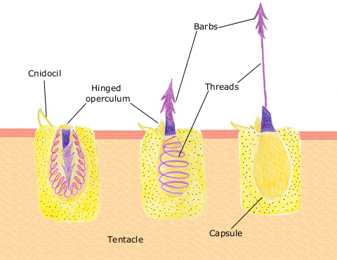
English: The diagram above shows the anatomy of a nematocyst cell and its “firing” sequence, from left to right. On the far left is a nematocyst inside its cellular capsule. The cell’s thread is coiled under pressure and wrapped around a stinging barb. When potential prey makes contact with the tentacles of a polyp, the nematocyst cell is stimulated. This causes a flap of tissue covering the nematocyst—the operculum—to fly open. The middle image shows the open operculum, the rapidly uncoiling thread and the emerging barb. On the far right is the fully extended cell. The barbs at the end of the nematocyst are designed to stick into the polyp’s victim and inject a poisonous liquid. When subdued, the polyp’s tentacles move the prey toward its mouth and the nematocysts recoil back into their capsules. |
| Датум | 11. април 2007. (првобитни датум слања) |
| Извор | Пребачено са en.wikipedia на Оставу. |
| Аутор | Датотеку је првобитно послао Spaully на енглески Википедија |
Лиценцирање
Ово дело је лиценцирано под условима Кријејтив комонс Делити под истим условима 1.0 лиценце.
|
This image is in the public domain because it contains materials that originally came from the U.S. National Oceanic and Atmospheric Administration, taken or made as part of an employee's official duties.
|
Првобитан дневник отпремања
Оригиналан опис странице је био овде. Сва наведена корисничка имена воде на en.wikipedia.
- 2007-04-11 17:10 Spaully 480×371×8 (194868 bytes) Modified from: http://www.oceanservice.noaa.gov/education/kits/corals/media/supp_coral01b.html {{Information |Description=Nematocyst discharge process. |Source= Modified from [http://www.oceanservice.noaa.gov/education/kits/corals/media/supp_coral01b.html
Историја датотеке
Кликните на датум/време да бисте видели тадашњу верзију датотеке.
| Датум/време | Минијатура | Димензије | Корисник | Коментар | |
|---|---|---|---|---|---|
| тренутна | 19:29, 13. октобар 2007. | | 480 × 371 (190 kB) | Alison | {{Information |Description===Description== The diagram above shows the anatomy of a nematocyst cell and its “firing” sequence, from left to right. On the far left is a nematocyst inside its cellular capsule. The cell’s thread is coiled under pressur |
Употреба датотеке
Нема страница које користе ову датотеку.
Глобална употреба датотеке
Други викији који користе ову датотеку:
- Употреба на ca.wikipedia.org
- Употреба на ceb.wikipedia.org
- Употреба на en.wikipedia.org
- Употреба на fr.wikipedia.org
- Употреба на hr.wikipedia.org
- Употреба на id.wikipedia.org
- Употреба на it.wikibooks.org
- Употреба на ja.wikipedia.org
- Употреба на lv.wikipedia.org
- Употреба на ms.wikipedia.org
- Употреба на my.wikipedia.org
- Употреба на pa.wikipedia.org
- Употреба на pt.wikipedia.org
- Употреба на simple.wikipedia.org
- Употреба на sv.wikipedia.org
- Употреба на te.wikipedia.org
- Употреба на th.wikipedia.org
- Употреба на vi.wikipedia.org