Покровни систем — разлика између измена
. ознака: везе до вишезначних одредница |
|||
| Ред 1: | Ред 1: | ||
{{short description|Систем органа који штити тело који се састоји од коже и њених додатака (укључујући косу, крљушти, перје, копита и нокте)}} |
|||
{{Infobox anatomy |
{{Infobox anatomy |
||
| Name = Покровни систем |
| Name = Покровни систем |
||
| Ред 31: | Ред 32: | ||
== Састав покровног система == |
== Састав покровног система == |
||
{{rut}} |
|||
Главни састојци покровног система су кожна мембрана (кожа), и њој придружене помоћне структуре (власи, нокти, [[егзокрине жлезде]]). |
Главни састојци покровног система су кожна мембрана (кожа), и њој придружене помоћне структуре (власи, нокти, [[егзокрине жлезде]]). Кожне жлезде су: знојне жлезде, лојне жлезде, млечне жлезде и ушне жлезде |
||
=== Skin === |
|||
Кожне жлезде су: |
|||
{{Main|Skin}} |
|||
* Знојне жлезде |
|||
The skin is one of the largest organs of the body. In humans, it accounts for about 12 to 15 percent of total body weight and covers 1.5-2m<sup>2</sup> of surface area.<ref>{{cite book |last1=Martini |first1=Frederic |last2=Nath |first2=Judi L. |title=Fundamentals of anatomy & physiology |date=2009 |publisher=Pearson/Benjamin Cummings |location=San Francisco |isbn=978-0321505897 |page=158 |edition=8th}}</ref> |
|||
* Лојне жлезде |
|||
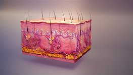
[[File:Integumentary system.jpg|thumb|263x263px|3D still showing human integumentary system.]] |
|||
* Млечне жлезде |
|||
* Ушне жлезде |
|||
The skin (integument) is a composite organ, made up of at least two major layers of tissue: the [[Epidermis (skin)|epidermis]] and the [[dermis]].<ref name="Kardong2019">{{cite book |last1=Kardong |first1=Kenneth V. |title=Vertebrates : comparative anatomy, function, evolution |date=2019 |location=New York, NY |isbn=978-1-259-70091-0 |pages=212–214 |edition=Eighth}}</ref> The epidermis is the outermost layer, providing the initial barrier to the external environment. It is separated from the dermis by the [[basement membrane]] ([[basal lamina]] and [[reticular lamina]]). The epidermis contains [[melanocyte]]s and gives color to the skin. The deepest layer of the epidermis also contains [[nerve ending]]s. Beneath this, the dermis comprises two sections, the papillary and reticular layers, and contains [[connective tissue]]s, vessels, glands, follicles, [[hair root]]s, sensory nerve endings, and muscular tissue.<ref name="aging skin">{{cite web |title=The Ageing Skin – Part 1 – Structure of Skin |url=http://pharmaxchange.info/press/2011/03/the-ageing-skin-part-1-structure-of-skin-and-introduction |website=pharmaxchange.info}}</ref> |
|||
Between the integument and the deep body musculature there is a transitional subcutaneous zone made up of very loose connective and [[adipose tissue]], the [[hypodermis]]. Substantial [[collagen]] bundles anchor the dermis to the hypodermis in a way that permits most areas of the skin to move freely over the deeper tissue layers.<ref>{{cite web|last=Pratt|first=Rebecca|title=Integument|url=http://www.anatomyone.com/a/integument/|work=AnatomyOne|publisher=Amirsys, Inc|access-date=2012-09-28}}</ref> |
|||
====Epidermis==== |
|||
{{Main|Epidermis}} |
|||
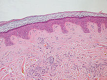
[[File:Normal Epidermis and Dermis with Intradermal Nevus 10x.JPG|thumb|left|Epidermis and dermis of human skin]] |
|||
The [[epidermis]] is the strong, superficial layer that serves as the first line of protection against the outer environment. The human epidermis is composed of [[Stratified squamous epithelium|stratified squamous epithelial cells]], which further break down into four to five layers: the [[stratum corneum]], [[stratum granulosum]], [[stratum spinosum]] and [[stratum basale]]. Where the skin is thicker, such as in the palms and soles, there is an extra layer of skin between the stratum corneum and the stratum granulosum, called the [[stratum lucidum]]. The epidermis is regenerated from the stem cells found in the basal layer that develop into the corneum. The epidermis itself is devoid of blood supply and draws its nutrition from its underlying dermis.<ref name="statpearls2"/> |
|||
Its main functions are protection, absorption of nutrients, and [[homeostasis]]. In structure, it consists of a keratinized stratified [[squamous epithelium]]; four types of cells: [[keratinocytes]], [[melanocytes]], [[Merkel cells]], and [[Langerhans cells]]. |
|||
The predominant cell type of the epidermis is the [[keratinocyte]], which produces [[keratin]], a fibrous protein that aids in skin protection, and is responsible for the formation of the epidermal water barrier by making and secreting [[lipid]]s.<ref name="statpearls">{{cite book |last1=Yousef |first1=Hani |last2=Alhajj |first2=Mandy |last3=Sharma |first3=Sandeep |title=StatPearls |publisher=StatPearls Publishing |url=https://www.ncbi.nlm.nih.gov/books/NBK470464/ |chapter=Anatomy, Skin (Integument), Epidermis}}</ref> <!--An overwhelming amount of keratin can cause disease by giving rise to eruptions from the skin that will protrude outwards and lead to infection.{{Citation needed|date=March 2017}}--> The majority of the skin on the human body is keratinized, with the exception of the lining of [[mucous membrane]]s, such as the inside of the mouth. Non-keratinized cells allow water to "stay" atop the structure. |
|||
The protein keratin stiffens epidermal tissue to form [[fingernail]]s. Nails grow from a thin area called the [[Matrix (nail)|nail matrix]] at an average of 1 mm per week. The [[lunula (anatomy)|lunula]] is the crescent-shape area at the base of the nail, lighter in color as it mixes with matrix cells. Only [[primate]]s have nails. In other vertebrates, the keratinizing system at the terminus of each digit produces claws or hooves.<ref name="Kardong2019"/> |
|||
The epidermis of vertebrates is surrounded by two kinds of coverings, which are produced by the epidermis itself. In [[fish]] and aquatic [[amphibian]]s, it is a thin mucus layer that is constantly being replaced. In terrestrial vertebrates, it is the [[stratum corneum]] (dead keratinized cells). The epidermis is, to some degree, glandular in all vertebrates, but more so in [[fish]] and [[amphibian]]s. Multicellular epidermal glands penetrate the dermis, where they are surrounded by blood capillaries that provide nutrients and, in the case of endocrine glands, transport their products.<ref>{{cite journal |last1=Quay |first1=Wilbur B. |title=Integument and the Environment Glandular Composition, Function, and Evolution |journal=Integrative and Comparative Biology |date=1 February 1972 |volume=12 |issue=1 |pages=95–108 |url=https://academic.oup.com/icb/article/12/1/95/2107657}}</ref> |
|||
{{Clear|left}} |
|||
====Dermis==== |
|||
{{Main|Dermis}} |
|||
The dermis is the underlying connective tissue layer that supports the epidermis. It is composed of dense irregular connective tissue and [[Loose connective tissue|areolar connective tissue]] such as a collagen with [[elastin]] arranged in a diffusely bundled and woven pattern. |
|||
The dermis has two layers: the papillary dermis and the reticular layer. The papillary layer is the superficial layer that forms finger-like projections into the epidermis (dermal papillae),<ref name="statpearls2">{{cite book |last1=Kim |first1=Joyce Y. |last2=Dao |first2=Harry |title=StatPearls |publisher=StatPearls Publishing |url=https://www.ncbi.nlm.nih.gov/books/NBK554386/ |chapter=Physiology, Integument}}</ref> and consists of highly vascularized, loose connective tissue. The reticular layer is the deep layer of the dermis and consists of the dense irregular connective tissue. These layers serve to give elasticity to the integument, allowing stretching and conferring flexibility, while also resisting distortions, wrinkling, and sagging.<ref name="aging skin"/> The dermal layer provides a site for the endings of blood vessels and nerves. Many [[chromatophores]] are also stored in this layer, as are the bases of integumental structures such as [[hair]], [[feathers]], and [[glands]]. |
|||
===Hypodermis=== |
|||
{{Main|Hypodermis}} |
|||
The hypodermis, otherwise known as the subcutaneous layer, is a layer beneath the skin. It invaginates into the dermis and is attached to the latter, immediately above it, by collagen and elastin fibers. It is essentially composed of a type of cell known as adipocytes, which are specialized in accumulating and storing fats. These cells are grouped together in lobules separated by connective tissue. |
|||
The hypodermis acts as an energy reserve. The fats contained in the adipocytes can be put back into circulation, via the venous route, during intense effort or when there is a lack of energy-providing substances, and are then transformed into energy. The hypodermis participates, passively at least, in thermoregulation since fat is a heat insulator. |
|||
==Functions== |
|||
The integumentary system has multiple roles in [[homeostasis|maintaining the body's equilibrium]]. All body systems work in an interconnected manner to maintain the internal conditions essential to the function of the body. The skin has an important job of protecting the body and acts as the body's first line of defense against infection, temperature change, and other challenges to homeostasis.<ref>{{MeSH name|Integumentary+System}}</ref><ref>{{cite book | last=Marieb | first=Elaine |author2=Hoehn, Katja | title=Human Anatomy & Physiology | url=https://archive.org/details/humananatomyphys00mari_4 | url-access=registration | publisher=[[Pearson Benjamin Cummings]] | year=2007 | edition=7th | page=[https://archive.org/details/humananatomyphys00mari_4/page/142 142]}}</ref> |
|||
Its main functions include: |
|||
*Protect the body's internal living [[Tissue (biology)|tissue]]s and organs |
|||
*Protect against invasion by [[Infection|infectious]] organisms |
|||
*Protect the body from [[dehydration]] |
|||
*Protect the body against [[Weather|abrupt changes]] in [[temperature]], maintain [[homeostasis]] |
|||
*Help [[Excretion|excrete]] waste materials through [[perspiration]] |
|||
*Act as a receptor for touch, pressure, pain, heat, and cold (see [[Somatosensory system]]) |
|||
*Protect the body against [[sunburn]]s by secreting melanin |
|||
*Generate [[vitamin D]] through exposure to [[ultraviolet]] [[light]] |
|||
*Store [[water]], [[fat]], glucose, vitamin D |
|||
*Maintenance of the body form |
|||
*Formation of new cells from stratum germinativum to repair minor injuries |
|||
*Protect from [[UV]] rays. |
|||
*Regulates body temperature |
|||
*It distinguishes, separates, and protects the organism from its surroundings. |
|||
Small-bodied invertebrates of aquatic or continually moist habitats [[Respiration (physiology)|respire]] using the outer layer (integument). This gas exchange system, where gases simply diffuse into and out of the [[interstitial fluid]], is called '''integumentary exchange'''. |
|||
== Дерматологија == |
== Дерматологија == |
||
| Ред 43: | Ред 96: | ||
== Референце == |
== Референце == |
||
{{reflist}} |
{{reflist|}} |
||
== Референце == |
|||
{{refbegin|30em}} |
|||
* {{cite book | last1=McGrath |first1=J.A. |last2=Eady|first2=R.A. |last3=Pope|first3=F.M. | title=Rook's Textbook of Dermatology | year=2004 | edition=7th | publisher=Blackwell Publishing | isbn=978-0-632-06429-8 | pages=3.1–3.6 }} |
|||
* {{Cite journal |last1=Varga |first1=Joseph F. A. |last2=Bui-Marinos |first2=Maxwell P. |last3=Katzenback |first3=Barbara A. |date=2019 |title=Frog Skin Innate Immune Defences: Sensing and Surviving Pathogens |journal=Frontiers in Immunology |volume=9 |page=3128 |doi=10.3389/fimmu.2018.03128 |pmid=30692997 |pmc=6339944 |issn=1664-3224|doi-access=free }} |
|||
* {{Cite journal |last1=Ferrie |first1=Gina M. |last2=Alford |first2=Vance C. |last3=Atkinson |first3=Jim |last4=Baitchman |first4=Eric |last5=Barber |first5=Diane |last6=Blaner |first6=William S. |last7=Crawshaw |first7=Graham |last8=Daneault |first8=Andy |last9=Dierenfeld |first9=Ellen |last10=Finke |first10=Mark |last11=Fleming |first11=Greg |date=2014 |title=Nutrition and Health in Amphibian Husbandry |journal=Zoo Biology |volume=33 |issue=6 |pages=485–501 |doi=10.1002/zoo.21180 |issn=0733-3188 |pmc=4685711 |pmid=25296396}} |
|||
* {{Cite journal |last=Varga |first=Joseph F. A. |last2=Bui-Marinos |first2=Maxwell P. |last3=Katzenback |first3=Barbara A. |date=2019 |title=Frog Skin Innate Immune Defences: Sensing and Surviving Pathogens |url=https://www.frontiersin.org/article/10.3389/fimmu.2018.03128 |journal=Frontiers in Immunology |volume=9 |doi=10.3389/fimmu.2018.03128/full |issn=1664-3224}} |
|||
* {{Cite web |last=Fisheries |first=NOAA |date=2022-05-03 |title=Fun Facts About Shocking Sharks {{!}} NOAA Fisheries |url=https://www.fisheries.noaa.gov/national/outreach-and-education/fun-facts-about-shocking-sharks |access-date=2022-05-11 |website=NOAA |language=en}} |
|||
* {{Cite journal |title=Shark and Ray Workbook 3-5 update 8-31 |url=https://www.floridaocean.org/sites/default/files/images/Shark%20and%20Ray%20Workbook%203-5%20update%208-31.pdf |journal=}} |
|||
* {{cite journal |last1=Toledo |first1=R.C. |last2=Jared |first2=C. |title=Cutaneous granular glands and amphibian venoms |journal=Comparative Biochemistry and Physiology Part A: Physiology |date=May 1995 |volume=111 |issue=1 |pages=1–29 |doi=10.1016/0300-9629(95)98515-I }} |
|||
* {{cite journal |last1=Dawson |first1=A. B. |title=The integument of necturus maculosus |journal=Journal of Morphology |date=December 1920 |volume=34 |issue=3 |pages=486–589 |doi=10.1002/jmor.1050340303 |s2cid=83534922 |url=https://books.google.com/books?id=UklQAQAAMAAJ&pg=PA487}} |
|||
* {{cite book |author=Romer, Alfred Sherwood|author2=Parsons, Thomas S.|year=1977 |title=The Vertebrate Body |publisher=Holt-Saunders International |location= Philadelphia|pages= 129–145|isbn= 978-0-03-910284-5}} |
|||
* {{Cite journal |title=714_1.tif |url=http://genesdev.cshlp.org/content/5/5/714.full.pdf |journal=}} |
|||
* {{cite journal |last1=Vassar |first1=R |last2=Fuchs |first2=E |title=Transgenic mice provide new insights into the role of TGF-alpha during epidermal development and differentiation |journal=Genes Dev |date=1 May 1991 |volume=5 |issue=5 |pages=714–727 |doi=10.1101/gad.5.5.714 |pmid=1709129 |doi-access=free }} |
|||
* {{Cite journal |last=Gilbert |first=Scott F. |date=2000 |title=Induction and Competence |url=https://www.ncbi.nlm.nih.gov/books/NBK9993/ |journal=Developmental Biology. 6th edition |language=en}} |
|||
* {{cite journal |last1=Stücker |first1=M. |last2=Struk |first2=A. |last3=Altmeyer |first3=P. |last4=Herde |first4=M. |last5=Baumgärtl |first5=H. |last6=Lübbers |first6=D. W. |title=The cutaneous uptake of atmospheric oxygen contributes significantly to the oxygen supply of human dermis and epidermis |journal=The Journal of Physiology |date=February 2002 |volume=538 |issue=3 |pages=985–994 |doi=10.1113/jphysiol.2001.013067 |pmid=11826181 |pmc=2290093 }} |
|||
* {{cite book|last=McCracken|first=Thomas|title=New Atlas of Human Anatomy|year=2000|publisher=Metro Books|location=China|isbn=978-1-58663-097-3|pages=1–240}} |
|||
* {{cite magazine |title=Camouflage |url=http://www.nationalgeographic.org/encyclopedia/camouflage/ |magazine=National Geographic |access-date=27 February 2017 |date=2011-08-25 |archive-url=https://web.archive.org/web/20170227232527/http://www.nationalgeographic.org/encyclopedia/camouflage/ |archive-date=27 February 2017 |url-status=live }} |
|||
{{refend}} |
|||
== Спољашње везе == |
== Спољашње везе == |
||
{{Commonscat|Integumentary system}} |
{{Commonscat|Integumentary system}} |
||
* {{Cite web |title=Pangolin Fact Sheet {{!}} Blog {{!}} Nature {{!}} PBS |url=https://www.pbs.org/wnet/nature/blog/pangolin-fact-sheet/ |access-date=2022-05-11 |website=Nature |language=en-US}} |
|||
{{нормативна контрола}} |
{{нормативна контрола}} |
||
Верзија на датум 29. мај 2022. у 18:09
| Називи и ознаке | |
|---|---|
| MeSH | D034582 |
| TA98 | A16.0.00.001 |
| TA2 | 7040 |
| TH | ТХ {{{2}}}.html HH3.12.00.0.00001 .{{{2}}}.{{{3}}} |
| FMA | 72979 |
| Анатомска терминологија | |
У зоологији, покровни систем је често највећи систем органа код животиња и састоји се од коже, длака, перја, крљушти, ноктију, кожних жлезда, и њихових излучевина (зној и лој).[1][2] Он одваја, штити, и обавештава животињу о стању у њеној околини.[3] Код малих бескичмењака који живе у води или влажним стаништима, покровни систем обавља и функцију дисања. Овај систем за размену гасова, којим гасови једноставно дифундују унутар и ван интерстицијске течности, се назива покровна размена.
У ботаници, реч покровни се односи на омотач неоплођеној јајашца.
Сама реч потиче од латинске речи integumentum чије је значење: прекрити.
Састав покровног система
Један корисник управо ради на овом чланку. Молимо остале кориснике да му допусте да заврши са радом. Ако имате коментаре и питања у вези са чланком, користите страницу за разговор.
Хвала на стрпљењу. Када радови буду завршени, овај шаблон ће бити уклоњен. Напомене
|
Главни састојци покровног система су кожна мембрана (кожа), и њој придружене помоћне структуре (власи, нокти, егзокрине жлезде). Кожне жлезде су: знојне жлезде, лојне жлезде, млечне жлезде и ушне жлезде
Skin
The skin is one of the largest organs of the body. In humans, it accounts for about 12 to 15 percent of total body weight and covers 1.5-2m2 of surface area.[4]

The skin (integument) is a composite organ, made up of at least two major layers of tissue: the epidermis and the dermis.[5] The epidermis is the outermost layer, providing the initial barrier to the external environment. It is separated from the dermis by the basement membrane (basal lamina and reticular lamina). The epidermis contains melanocytes and gives color to the skin. The deepest layer of the epidermis also contains nerve endings. Beneath this, the dermis comprises two sections, the papillary and reticular layers, and contains connective tissues, vessels, glands, follicles, hair roots, sensory nerve endings, and muscular tissue.[6]
Between the integument and the deep body musculature there is a transitional subcutaneous zone made up of very loose connective and adipose tissue, the hypodermis. Substantial collagen bundles anchor the dermis to the hypodermis in a way that permits most areas of the skin to move freely over the deeper tissue layers.[7]
Epidermis

The epidermis is the strong, superficial layer that serves as the first line of protection against the outer environment. The human epidermis is composed of stratified squamous epithelial cells, which further break down into four to five layers: the stratum corneum, stratum granulosum, stratum spinosum and stratum basale. Where the skin is thicker, such as in the palms and soles, there is an extra layer of skin between the stratum corneum and the stratum granulosum, called the stratum lucidum. The epidermis is regenerated from the stem cells found in the basal layer that develop into the corneum. The epidermis itself is devoid of blood supply and draws its nutrition from its underlying dermis.[8]
Its main functions are protection, absorption of nutrients, and homeostasis. In structure, it consists of a keratinized stratified squamous epithelium; four types of cells: keratinocytes, melanocytes, Merkel cells, and Langerhans cells.
The predominant cell type of the epidermis is the keratinocyte, which produces keratin, a fibrous protein that aids in skin protection, and is responsible for the formation of the epidermal water barrier by making and secreting lipids.[9] The majority of the skin on the human body is keratinized, with the exception of the lining of mucous membranes, such as the inside of the mouth. Non-keratinized cells allow water to "stay" atop the structure.
The protein keratin stiffens epidermal tissue to form fingernails. Nails grow from a thin area called the nail matrix at an average of 1 mm per week. The lunula is the crescent-shape area at the base of the nail, lighter in color as it mixes with matrix cells. Only primates have nails. In other vertebrates, the keratinizing system at the terminus of each digit produces claws or hooves.[5]
The epidermis of vertebrates is surrounded by two kinds of coverings, which are produced by the epidermis itself. In fish and aquatic amphibians, it is a thin mucus layer that is constantly being replaced. In terrestrial vertebrates, it is the stratum corneum (dead keratinized cells). The epidermis is, to some degree, glandular in all vertebrates, but more so in fish and amphibians. Multicellular epidermal glands penetrate the dermis, where they are surrounded by blood capillaries that provide nutrients and, in the case of endocrine glands, transport their products.[10]
Dermis
The dermis is the underlying connective tissue layer that supports the epidermis. It is composed of dense irregular connective tissue and areolar connective tissue such as a collagen with elastin arranged in a diffusely bundled and woven pattern.
The dermis has two layers: the papillary dermis and the reticular layer. The papillary layer is the superficial layer that forms finger-like projections into the epidermis (dermal papillae),[8] and consists of highly vascularized, loose connective tissue. The reticular layer is the deep layer of the dermis and consists of the dense irregular connective tissue. These layers serve to give elasticity to the integument, allowing stretching and conferring flexibility, while also resisting distortions, wrinkling, and sagging.[6] The dermal layer provides a site for the endings of blood vessels and nerves. Many chromatophores are also stored in this layer, as are the bases of integumental structures such as hair, feathers, and glands.
Hypodermis
The hypodermis, otherwise known as the subcutaneous layer, is a layer beneath the skin. It invaginates into the dermis and is attached to the latter, immediately above it, by collagen and elastin fibers. It is essentially composed of a type of cell known as adipocytes, which are specialized in accumulating and storing fats. These cells are grouped together in lobules separated by connective tissue.
The hypodermis acts as an energy reserve. The fats contained in the adipocytes can be put back into circulation, via the venous route, during intense effort or when there is a lack of energy-providing substances, and are then transformed into energy. The hypodermis participates, passively at least, in thermoregulation since fat is a heat insulator.
Functions
The integumentary system has multiple roles in maintaining the body's equilibrium. All body systems work in an interconnected manner to maintain the internal conditions essential to the function of the body. The skin has an important job of protecting the body and acts as the body's first line of defense against infection, temperature change, and other challenges to homeostasis.[11][12]
Its main functions include:
- Protect the body's internal living tissues and organs
- Protect against invasion by infectious organisms
- Protect the body from dehydration
- Protect the body against abrupt changes in temperature, maintain homeostasis
- Help excrete waste materials through perspiration
- Act as a receptor for touch, pressure, pain, heat, and cold (see Somatosensory system)
- Protect the body against sunburns by secreting melanin
- Generate vitamin D through exposure to ultraviolet light
- Store water, fat, glucose, vitamin D
- Maintenance of the body form
- Formation of new cells from stratum germinativum to repair minor injuries
- Protect from UV rays.
- Regulates body temperature
- It distinguishes, separates, and protects the organism from its surroundings.
Small-bodied invertebrates of aquatic or continually moist habitats respire using the outer layer (integument). This gas exchange system, where gases simply diffuse into and out of the interstitial fluid, is called integumentary exchange.
Дерматологија
Дерматологија је грана медицине која се бави покровним системом. Како је кожа најуочљивији орган, то њен изглед или симптоми дају информације за важне закључке везане за кожне болести или чак везане и за болести других органа, попут јетре. Кожа је такојже најрањивији орган због своје изложености зрачењу, физичким повредама, инфекцијама и штетним хемикалијама.
Референце
- ^ Integumentary+System на US National Library of Medicine Medical Subject Headings (MeSH)
- ^ Marieb, Elaine; Katja Hoehn (2007). Human Anatomy & Physiology (7th изд.). Pearson Benjamin Cummings. стр. 142.
- ^ „The Integumentary System”. Encyclopedia.com. Приступљено 3. 6. 2013.
- ^ Martini, Frederic; Nath, Judi L. (2009). Fundamentals of anatomy & physiology (8th изд.). San Francisco: Pearson/Benjamin Cummings. стр. 158. ISBN 978-0321505897.
- ^ а б Kardong, Kenneth V. (2019). Vertebrates : comparative anatomy, function, evolution (Eighth изд.). New York, NY. стр. 212—214. ISBN 978-1-259-70091-0.
- ^ а б „The Ageing Skin – Part 1 – Structure of Skin”. pharmaxchange.info.
- ^ Pratt, Rebecca. „Integument”. AnatomyOne. Amirsys, Inc. Приступљено 2012-09-28.
- ^ а б Kim, Joyce Y.; Dao, Harry. „Physiology, Integument”. StatPearls. StatPearls Publishing.
- ^ Yousef, Hani; Alhajj, Mandy; Sharma, Sandeep. „Anatomy, Skin (Integument), Epidermis”. StatPearls. StatPearls Publishing.
- ^ Quay, Wilbur B. (1. 2. 1972). „Integument and the Environment Glandular Composition, Function, and Evolution”. Integrative and Comparative Biology. 12 (1): 95—108.
- ^ Integumentary+System на US National Library of Medicine Medical Subject Headings (MeSH)
- ^ Marieb, Elaine; Hoehn, Katja (2007). Human Anatomy & Physiology
 (7th изд.). Pearson Benjamin Cummings. стр. 142.
Референце
- McGrath, J.A.; Eady, R.A.; Pope, F.M. (2004). Rook's Textbook of Dermatology (7th изд.). Blackwell Publishing. стр. 3.1—3.6. ISBN 978-0-632-06429-8.
- Varga, Joseph F. A.; Bui-Marinos, Maxwell P.; Katzenback, Barbara A. (2019). „Frog Skin Innate Immune Defences: Sensing and Surviving Pathogens”. Frontiers in Immunology. 9: 3128. ISSN 1664-3224. PMC 6339944
 . PMID 30692997. doi:10.3389/fimmu.2018.03128
  .
. - Ferrie, Gina M.; Alford, Vance C.; Atkinson, Jim; Baitchman, Eric; Barber, Diane; Blaner, William S.; Crawshaw, Graham; Daneault, Andy; Dierenfeld, Ellen; Finke, Mark; Fleming, Greg (2014). „Nutrition and Health in Amphibian Husbandry”. Zoo Biology. 33 (6): 485—501. ISSN 0733-3188. PMC 4685711
 . PMID 25296396. doi:10.1002/zoo.21180.
 - Varga, Joseph F. A.; Bui-Marinos, Maxwell P.; Katzenback, Barbara A. (2019). „Frog Skin Innate Immune Defences: Sensing and Surviving Pathogens”. Frontiers in Immunology. 9. ISSN 1664-3224. doi:10.3389/fimmu.2018.03128/full.
- Fisheries, NOAA (2022-05-03). „Fun Facts About Shocking Sharks | NOAA Fisheries”. NOAA (на језику: енглески). Приступљено 2022-05-11.
- „Shark and Ray Workbook 3-5 update 8-31” (PDF).
- Toledo, R.C.; Jared, C. (мај 1995). „Cutaneous granular glands and amphibian venoms”. Comparative Biochemistry and Physiology Part A: Physiology. 111 (1): 1—29. doi:10.1016/0300-9629(95)98515-I.
- Dawson, A. B. (децембар 1920). „The integument of necturus maculosus”. Journal of Morphology. 34 (3): 486—589. S2CID 83534922. doi:10.1002/jmor.1050340303.
- Romer, Alfred Sherwood; Parsons, Thomas S. (1977). The Vertebrate Body. Philadelphia: Holt-Saunders International. стр. 129—145. ISBN 978-0-03-910284-5.
- „714_1.tif” (PDF).
- Vassar, R; Fuchs, E (1. 5. 1991). „Transgenic mice provide new insights into the role of TGF-alpha during epidermal development and differentiation”. Genes Dev. 5 (5): 714—727. PMID 1709129. doi:10.1101/gad.5.5.714
 .
. - Gilbert, Scott F. (2000). „Induction and Competence”. Developmental Biology. 6th edition (на језику: енглески).
- Stücker, M.; Struk, A.; Altmeyer, P.; Herde, M.; Baumgärtl, H.; Lübbers, D. W. (фебруар 2002). „The cutaneous uptake of atmospheric oxygen contributes significantly to the oxygen supply of human dermis and epidermis”. The Journal of Physiology. 538 (3): 985—994. PMC 2290093
 . PMID 11826181. doi:10.1113/jphysiol.2001.013067.
 - McCracken, Thomas (2000). New Atlas of Human Anatomy. China: Metro Books. стр. 1—240. ISBN 978-1-58663-097-3.
- „Camouflage”. National Geographic. 2011-08-25. Архивирано из оригинала 27. 2. 2017. г. Приступљено 27. 2. 2017.
Спољашње везе
- „Pangolin Fact Sheet | Blog | Nature | PBS”. Nature (на језику: енглески). Приступљено 2022-05-11.
