Гуштерача — разлика између измена
Add 1 book for Википедија:Проверљивост (20230201)) #IABot (v2.0.9.3) (GreenC bot |
. ознака: везе до вишезначних одредница |
||
| Ред 1: | Ред 1: | ||
{{rut}} |
|||
[[Датотека:Gray 1100 Pancreatic duct.png|мини|десно|200п|Гуштерача]] |
|||
{{Infobox anatomy |
|||
'''Гуштерача''', позната и под називом '''панкреас''', је веома важна [[жлезда]] у систему органа за варење.<ref name="MEDSCAPE">{{cite web|last=Khan|first=Ali Nawaz|title=Chronic Pancreatitis Imaging|url=http://emedicine.medscape.com/article/371772-overview|work=Medscape|accessdate=05. 01. 2014}}</ref> Дугачка је отприлике 15 -{cm}-, дугуљастог облика и пљосната.<ref>{{cite web|title=Cancer of the Pancreas|url=http://www.nhs.uk/conditions/Cancer-of-the-pancreas/Pages/Introduction.aspx|website=NHS|accessdate=05. 11. 2014}}</ref> |
|||
| Name = Гуштерача |
|||
| Latin = Pancreas |
|||
| Greek = Πάνκρεας (Pánkreas) |
|||
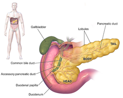
| Image = Blausen_0699_PancreasAnatomy2.png |
|||
| Caption = Anatomy of the pancreas |
|||
| Width = |
|||
| Precursor = [[Pancreatic bud]]s |
|||
| System = [[Digestive system]] and [[endocrine system]] |
|||
| Artery = [[Inferior pancreaticoduodenal artery]], [[anterior superior pancreaticoduodenal artery]], [[posterior superior pancreaticoduodenal artery]], [[splenic artery]] |
|||
| Vein = [[Pancreaticoduodenal veins]], [[pancreatic veins]] |
|||
| Nerve = [[Pancreatic plexus]], [[celiac ganglia]], [[vagus nerve]]<ref>{{cite book| title= Essentials of Human Physiology| first= Thomas M. |last= Nosek| chapter=Section 6/6ch2/s6ch2_30 |chapter-url=http://humanphysiology.tuars.com/program/section6/6ch2/s6ch2_30.htm |archive-url=https://web.archive.org/web/20160324124828/http://humanphysiology.tuars.com/program/section6/6ch2/s6ch2_30.htm|archive-date=2016-03-24}}</ref> |
|||
| Lymph = [[Splenic lymph nodes]], [[celiac lymph nodes]] and [[superior mesenteric lymph nodes]] |
|||
| Pronunciation = {{IPAc-en|ˈ|p|æ|ŋ|k|r|i|ə|s}} |
|||
}} |
|||
'''Гуштерача''', позната и под називом '''панкреас''', је веома важна [[жлезда]] у систему [[digestive system|органа за варење]] and [[endocrine system]] of [[vertebrate]]s.<ref name="MEDSCAPE">{{cite web|last=Khan|first=Ali Nawaz|title=Chronic Pancreatitis Imaging|url=http://emedicine.medscape.com/article/371772-overview|work=Medscape|accessdate=05. 01. 2014}}</ref> Дугачка је отприлике 15 -{cm}-, дугуљастог облика и пљосната.<ref>{{cite web|title=Cancer of the Pancreas|url=http://www.nhs.uk/conditions/Cancer-of-the-pancreas/Pages/Introduction.aspx|website=NHS|accessdate=05. 11. 2014}}</ref> In humans, it is located in the [[abdominal cavity|abdomen]] behind the [[stomach]] and functions as a [[gland]]. The pancreas is a mixed or [[heterocrine gland]], i.e. it has both an [[endocrine]] and a digestive [[exocrine]] function.<ref>{{Cite journal|last1=Gyr|first1=K.|last2=Beglinger|first2=C.|last3=Stalder|first3=G. A.|date=1985-09-21|title=[Interaction of the endo- and exocrine pancreas]|url=https://pubmed.ncbi.nlm.nih.gov/2865807/|journal=Schweizerische Medizinische Wochenschrift|volume=115|issue=38|pages=1299–1306|issn=0036-7672|pmid=2865807}}</ref> 99% of the pancreas is exocrine and 1% is endocrine.<ref>{{Cite web|title=Pancreas Gland - Endocrine System|url=https://www.innerbody.com/image/endo03.html|access-date=2021-06-12|website=Innerbody|language=en}}</ref>{{sfn|Beger's|2018|pp=124}}<ref>{{Cite web|title=Endocrine Pancreas - an overview ScienceDirect Topics|url=https://www.sciencedirect.com/topics/medicine-and-dentistry/endocrine-pancreas|access-date=2021-06-12|website=www.sciencedirect.com}}</ref><ref>{{Cite journal|last1=Henderson|first1=J. R.|last2=Daniel|first2=P. M.|last3=Fraser|first3=P. A.|date=February 1981|title=The pancreas as a single organ: the influence of the endocrine upon the exocrine part of the gland|journal=Gut|volume=22|issue=2|pages=158–167|doi=10.1136/gut.22.2.158|issn=0017-5749|pmc=1419227|pmid=6111521}}</ref> As an [[endocrine gland]], it functions mostly to regulate [[blood sugar level]]s, secreting the [[hormone]]s [[insulin]], [[glucagon]], [[somatostatin]], and [[pancreatic polypeptide]]. As a part of the digestive system, it functions as an [[exocrine gland]] secreting [[pancreatic juice]] into the [[duodenum]] through the [[pancreatic duct]]. This juice contains [[bicarbonate]], which neutralizes [[acid]] entering the duodenum from the stomach; and [[digestive enzyme]]s, which break down [[carbohydrates]], [[protein]]s, and [[lipids|fats]] in food entering the duodenum from the stomach. |
|||
== Грађа == |
== Грађа == |
||
| Ред 78: | Ред 94: | ||
#* ектопички панкреас |
#* ектопички панкреас |
||
# Дијабетес тип један (''-{diabetes mellitus typ I}-'') |
# Дијабетес тип један (''-{diabetes mellitus typ I}-'') |
||
== Историја == |
|||
The pancreas was first identified by [[Herophilus]] (335–280 BC), a [[Greeks|Greek]] [[anatomist]] and [[surgery|surgeon]].<ref>{{Cite book | url = https://books.google.com/books?id=rlLPBgAAQBAJ | title = History of the Pancreas: Mysteries of a Hidden Organ | last1 = Howard | first1 = John M. | last2 = Hess | first2 = Walter | date = 2012 | publisher = Springer Science & Business Media | isbn = 978-1461505556 | page = 24 | language = en }}</ref> A few hundred years later, [[Rufus of Ephesus]], another Greek anatomist, gave the pancreas its name. Etymologically, the term "pancreas", a modern [[Latin]] adaptation of [[Greek language|Greek]] πάγκρεας,<ref name="O'Brien">{{cite book| first = Terry | last = O'Brien |title=A2Z Book of word Origins|url=https://books.google.com/books?id=Rgzh83cChl0C&pg=PT86|publisher=Rupa Publications|isbn=978-8129118097|page=86|year=2015}}</ref> [πᾶν ("all", "whole"), and κρέας ("flesh")],<ref>{{cite web |last=Harper |first=Douglas |title=Pancreas |work=Online Etymology Dictionary |url=http://www.etymonline.com/index.php?term=pancreas |access-date=2007-04-04}}</ref> originally means [[sweetbread]],<ref name="Green2008">{{cite book| first = Tamara M. | last = Green |title=The Greek and Latin Roots of English|url=https://books.google.com/books?id=6gknyzq0uD8C&pg=PA176|year=2008|publisher=Rowman & Littlefield|isbn=978-0742547803|page=176}}</ref> although literally meaning all-flesh, presumably because of its fleshy consistency. It was only in 1889 when [[Oskar Minkowski]] discovered that removing the pancreas from a dog caused it to become diabetic.<ref name="Karamanou2016">{{cite journal |last1=Karamanou |first1=M |last2=Protogerou |first2=A |last3=Tsoucalas |first3=G |last4=Androutsos |first4=G |last5=Poulakou-Rebelakou |first5=E |title=Milestones in the history of diabetes mellitus: The main contributors. |journal=World Journal of Diabetes |date=10 January 2016 |volume=7 |issue=1 |pages=1–7 |doi=10.4239/wjd.v7.i1.1 |pmid=26788261|pmc=4707300 }}</ref> Insulin was later isolated from pancreatic islets by [[Frederick Banting]] and [[Charles Herbert Best]] in 1921.<ref name=Karamanou2016 /> |
|||
The way the tissue of the pancreas has been viewed has also changed. Previously, it was viewed using simple [[staining]] methods such as [[H&E stain]]s. Now, [[immunohistochemistry]] can be used to more easily differentiate cell types. This involves visible antibodies to the products of certain cell types, and helps identify with greater ease cell types such as alpha and beta cells.{{sfn|Wheater's Histology|2013|pp=332-333}} |
|||
== Референце == |
== Референце == |
||
| Ред 83: | Ред 105: | ||
== Литература == |
== Литература == |
||
{{refbegin|30em}} |
|||
* {{Cite book| ref=harv|editor-first=Barbara |editor-last=Young|title=Wheater's functional histology : a text and colour atlas| url=https://archive.org/details/wheatersfunction00youn|year=2006|publisher=Churchill Livingstone/Elsevier|isbn=978-0443068508|edition=5th|pages=[https://archive.org/details/wheatersfunction00youn/page/n672 299]-301}} |
* {{Cite book| ref=harv|editor-first=Barbara |editor-last=Young|title=Wheater's functional histology : a text and colour atlas| url=https://archive.org/details/wheatersfunction00youn|year=2006|publisher=Churchill Livingstone/Elsevier|isbn=978-0443068508|edition=5th|pages=[https://archive.org/details/wheatersfunction00youn/page/n672 299]-301}} |
||
* {{Cite book| ref=harv|last=Drake|first=Richard L.|title=Gray's anatomy for students| url=https://archive.org/details/graysanatomyfors00unse|year=2005|publisher=Elsevier/Churchill Livingstone|location=Philadelphia|isbn=978-0808923060|last2=Vogl|first2=Wayne|author3=Tibbitts, Adam W. M. Mitchell |last4=Richard|first4=illustrations by|last5=Richardson|first5=Paul|pages=[https://archive.org/details/graysanatomyfors00unse/page/288 288]-90, 297, 303}} |
* {{Cite book| ref=harv|last=Drake|first=Richard L.|title=Gray's anatomy for students| url=https://archive.org/details/graysanatomyfors00unse|year=2005|publisher=Elsevier/Churchill Livingstone|location=Philadelphia|isbn=978-0808923060|last2=Vogl|first2=Wayne|author3=Tibbitts, Adam W. M. Mitchell |last4=Richard|first4=illustrations by|last5=Richardson|first5=Paul|pages=[https://archive.org/details/graysanatomyfors00unse/page/288 288]-90, 297, 303}} |
||
* {{Cite book|title=Ganong's review of medical physiology|author1=Barrett, Kim E.|others=Barman, Susan M.,, Brooks, Heddwen L., Yuan, Jason X.-J.|isbn=9781260122404|edition=26th|location=New York|oclc=1076268769|year=2019|ref={{harvid|Ganong's Physiology|2019}}}} |
|||
* {{Cite book |editor=Beger HG|title=The pancreas : an integrated textbook of basic science, medicine, and surgery|date=2018|isbn=978-1-119-18841-4|location=Hoboken, NJ|oclc=1065547789|edition=third|ref={{harvid|Beger's|2018}}}} |
|||
* {{cite book |last1=Kasper |first1=Dennis |last2=Fauci |first2=Anthony |last3=Hauser |first3=Stephen |last4=Longo |first4=Dan |last5=Jameson |first5=J. |last6=Loscalzo |first6=Joseph |title=Harrison's Principles of Internal Medicine |date=2015 |publisher=McGraw-Hill Professional |isbn=9780071802154 |edition= 19|ref={{harvid|Harrison's|2015}}}} |
|||
* {{cite book |editor-last1=Ralston |editor-first1=Stuart H. |editor-last2=Penman |editor-first2=Ian D. |editor-last3=Strachan |editor-first3=Mark W. |editor-last4=Hobson |editor-first4=Richard P. |title=Davidson's principles and practice of medicine |date=2018 |publisher=Elsevier |isbn=978-0-7020-7028-0 |edition= 23rd|ref={{harvid|Davidson's|2018}}}} |
|||
* {{cite book |editor=Standring, Susan|editor2=Borley, Neil R.|title=Gray's anatomy : the anatomical basis of clinical practice|date=2008|publisher=Churchill Livingstone|location=London|isbn=978-0-8089-2371-8|edition= 40th|ref={{harvid|Gray's|2008}}|display-editors=etal}} |
|||
* {{Cite book |editor=Standring, Susan|title=Gray's anatomy : the anatomical basis of clinical practice|isbn=9780702052309|edition=41st|location=Philadelphia|oclc=920806541|year=2016|ref={{harvid|Gray's|2016}}}} |
|||
{{refend}} |
|||
== Спољашње везе == |
== Спољашње везе == |
||
Верзија на датум 25. фебруар 2023. у 19:37
Један корисник управо ради на овом чланку. Молимо остале кориснике да му допусте да заврши са радом. Ако имате коментаре и питања у вези са чланком, користите страницу за разговор.
Хвала на стрпљењу. Када радови буду завршени, овај шаблон ће бити уклоњен. Напомене
|
Гуштерача, позната и под називом панкреас, је веома важна жлезда у систему органа за варење and endocrine system of vertebrates.[2] Дугачка је отприлике 15 cm, дугуљастог облика и пљосната.[3] In humans, it is located in the abdomen behind the stomach and functions as a gland. The pancreas is a mixed or heterocrine gland, i.e. it has both an endocrine and a digestive exocrine function.[4] 99% of the pancreas is exocrine and 1% is endocrine.[5][6][7][8] As an endocrine gland, it functions mostly to regulate blood sugar levels, secreting the hormones insulin, glucagon, somatostatin, and pancreatic polypeptide. As a part of the digestive system, it functions as an exocrine gland secreting pancreatic juice into the duodenum through the pancreatic duct. This juice contains bicarbonate, which neutralizes acid entering the duodenum from the stomach; and digestive enzymes, which break down carbohydrates, proteins, and fats in food entering the duodenum from the stomach.
Грађа
Састоји се од:[9]
- главе (caput pancreatis) са продужетком (processus uncinatus)
- средњег дела, тела (corpus pancreatis) и
- дугачког, суженог дела, репа (cauda pancreatis).
Тежине је између 50 и 150 g. Добро је заштићена и смештена дубоко у горњем делу стомака, ретроперитонеално, у висини другог лумбалног пршљена иза желуца и дванаестопалачног црева, непосредно испред кичме.
Ембрионални развој
Панкреас настаје из једног трбушног вентралног и једног леђног дорзалног дела. У току развоја прелази вентрални део заједно са делом жучног пута (ductus choledochus) на дорзалну страну и сједињује се са дорзалним делом.
Функција
Гуштерача обавља двоструку улогу:[10]
- излучује сокове потребне за прераду хране у цревима, који се мешају са жучи из јетре. То је егзокрина функција гуштераче;
- у крв лучи хормоне, који делују у другим деловима тела, што представља ендокрину функцију гуштераче.
Егзокрина функција се огледа у лучењу панкреасног сока који се преко Вирсунговог изводног канала излива у нисходни део дванаестопалачног црева. Поред овог главног изводног канала постоји још један помоћни, Санторинијев изводни канал. Вирсунгов канал се најчешће сједињује са главним жучним путем (Ducus choledochus). На месту где се ови изводни канали уливају у силазни део дуоденума постоји мала избочина (papilla duodeni), око које се налазе мишићна влакна, која граде Одијев сфинктер, мишић који регулише ослобађање жучи и панкреасног сока.
Дневно панкреас човека излучи око 2000 ml сока. Основни састојци панкреасног сока су ензими за варење хране и најважнији међу њима су:
- трипсин,
- химотрипсин,
- липаза и
- амилаза.
Егзокрини панкреас секретује још и фосфолипазу, еластазу и рибонуклеазу.
Трипсин и химотрипсин учествују у варењу протеина па се излучују у неактивном облику, као трипсиноген и химотрипсиноген, да би се активирали тек када доспеју у танко црево. То су такозвани зимогени ензими. Прво се помоћу цревне ентерокиназе активира почетна, мала количина трипсиногена па затим он активира преосталу количину како трипсиногена тако и химотрипсиногена. Липаза се, такође, излучује у неактивног облику, а активира се тек у танком цреву помоћу жучи (ствара се у јетри и преко жучних канала доспава у дуоденум). Липаза делује само на емулговане масти, а да би се оне емулговале неопходна је жуч. Амилаза разлаже скроб.
Егзокрина функција и њена регулација
Постоје три фазе у регулацији лучења панкреасног сока:
- Цефалична фаза:
- мирис хране
- укус хране
- изглед јела - визуелна компонента изазива лучење панкреасног сока преко дејства нерва вагуса али хормон холецистокинин утиче на ове процесе
- Желудачна фаза - ширење зида желуца под утицајем хране активира вагус, а такође и покреће перисталтичку реакцију (перисталтика) уз помоћ безусловних рефлекса.
- Цревна фаза
Лучење панкреасног сока врши се само после доспевања хране у дуоденум, а свој максимум достиже три сата по уношењу хране. То лучење је под утицајем хормона секретина кога стварају жлезде танког црева када садржај из желуца доспе у њега. Секретин из танког црева крвљу доспева у панкреас где изазива лучење панкреасног сока богатог водом и бикарбонатима. На лучење овог сока утиче још један хормон, холецистокинин-панкреозимин. Овај ензим релаксира Одијев сфинктер, што омогућава секрецију панкреасног сока и жучи, али и стимулише панкреас на лучење сока богатог ензимима. Уједно холецистокинин инхибира активност желуца, што, омогућава да се храна која је доспела из желуца у дуоденум адекватно свари.
Поред хормоналне регулације, панкреасни сок се лучи и деловањем безусловних рефлекса (поглед на храну, мирис хране изазивају ту секрецију).
Ендокрина функција и њена регулација

Ендокрина функција панкреаса огледа се у раду ендокриних ћелија груписаних у тзв. Лангерхансова острвца. Ова острвца су разбацана по егзокрином панкреасу и смештена су између његових мешкова. Највише их има у репу панкреаса.
Ова острвца се састоје из три типа ћелија: алфа, бета и делта. Бета ћелије излучују хормон инсулин (лат. insula - острво), док алфа излучују глукагон. Инсулин и глукагон су са антагонистичким дејством: док инсулин смањује концентрацију глукозе у крви, глукагон је повећава.
Делта ћелије луче хормон соматостатин који инхибира секрецију осталих хормона. Такође панкреас секретује још један хормон амилин, који регулише брзину ресорпције у гастроинтестиналоом систему.
Повећана концентрација глукозе (шећера) у крви доводи до појачаног лучења инсулина па он смањује ниво шећера у крви. Када се ниво глукозе смањи на нормалу, смањује се и лучење инсулина. Међутим, када се инсулин недовољно излучује или постоји резистенција на њега долази до нагомилавања глукозе у крви, тј. хипергликемије (вредности веће од 6,66 mmol/l) што доводи до шећерне болести (diabetes mellitus).
Поремећаји функције панкреаса
- Акутно запаљење панкреаса (pankreatitis akuta. Постоје различите форме:
- серозни,
- егзудативни,
- хеморагични,
- некротични
- Хронично запаљење панкраса-pankreatitis chronica
- Рак, карцином панкреаса
- Аденом ендокриног панкреса:
- инсулином
- глукагоном
- гастрином (Синдром Золингер-Елисон)
- ВИП-ом, БИП-базоактивни интестинални пептид
- Соматостатином
- Развојне аномалије панкреаса:
- подељени панкреас (pankreas divisum)
- прстенасти панкреас (pankreas anulare)
- ектопички панкреас
- Дијабетес тип један (diabetes mellitus typ I)
Историја
The pancreas was first identified by Herophilus (335–280 BC), a Greek anatomist and surgeon.[11] A few hundred years later, Rufus of Ephesus, another Greek anatomist, gave the pancreas its name. Etymologically, the term "pancreas", a modern Latin adaptation of Greek πάγκρεας,[12] [πᾶν ("all", "whole"), and κρέας ("flesh")],[13] originally means sweetbread,[14] although literally meaning all-flesh, presumably because of its fleshy consistency. It was only in 1889 when Oskar Minkowski discovered that removing the pancreas from a dog caused it to become diabetic.[15] Insulin was later isolated from pancreatic islets by Frederick Banting and Charles Herbert Best in 1921.[15]
The way the tissue of the pancreas has been viewed has also changed. Previously, it was viewed using simple staining methods such as H&E stains. Now, immunohistochemistry can be used to more easily differentiate cell types. This involves visible antibodies to the products of certain cell types, and helps identify with greater ease cell types such as alpha and beta cells.[16]
Референце
- ^ Nosek, Thomas M. „Section 6/6ch2/s6ch2_30”. Essentials of Human Physiology. Архивирано из оригинала 2016-03-24. г.
- ^ Khan, Ali Nawaz. „Chronic Pancreatitis Imaging”. Medscape. Приступљено 05. 01. 2014.
- ^ „Cancer of the Pancreas”. NHS. Приступљено 05. 11. 2014.
- ^ Gyr, K.; Beglinger, C.; Stalder, G. A. (1985-09-21). „[Interaction of the endo- and exocrine pancreas]”. Schweizerische Medizinische Wochenschrift. 115 (38): 1299—1306. ISSN 0036-7672. PMID 2865807.
- ^ „Pancreas Gland - Endocrine System”. Innerbody (на језику: енглески). Приступљено 2021-06-12.
- ^ Beger's 2018, стр. 124.
- ^ „Endocrine Pancreas - an overview ScienceDirect Topics”. www.sciencedirect.com. Приступљено 2021-06-12.
- ^ Henderson, J. R.; Daniel, P. M.; Fraser, P. A. (фебруар 1981). „The pancreas as a single organ: the influence of the endocrine upon the exocrine part of the gland”. Gut. 22 (2): 158—167. ISSN 0017-5749. PMC 1419227
 . PMID 6111521. doi:10.1136/gut.22.2.158.
- ^ Drake 2005, стр. 288–90, 297, 303
- ^ Young, Barbara, ур. (2006). Wheater's functional histology : a text and colour atlas (5th изд.). Churchill Livingstone/Elsevier. стр. 299-301. ISBN 978-0443068508.
- ^ Howard, John M.; Hess, Walter (2012). History of the Pancreas: Mysteries of a Hidden Organ (на језику: енглески). Springer Science & Business Media. стр. 24. ISBN 978-1461505556.
- ^ O'Brien, Terry (2015). A2Z Book of word Origins. Rupa Publications. стр. 86. ISBN 978-8129118097.
- ^ Harper, Douglas. „Pancreas”. Online Etymology Dictionary. Приступљено 2007-04-04.
- ^ Green, Tamara M. (2008). The Greek and Latin Roots of English. Rowman & Littlefield. стр. 176. ISBN 978-0742547803.
- ^ а б Karamanou, M; Protogerou, A; Tsoucalas, G; Androutsos, G; Poulakou-Rebelakou, E (10. 1. 2016). „Milestones in the history of diabetes mellitus: The main contributors.”. World Journal of Diabetes. 7 (1): 1—7. PMC 4707300
 . PMID 26788261. doi:10.4239/wjd.v7.i1.1.
- ^ Wheater's Histology 2013, стр. 332–333.
Литература
- Young, Barbara, ур. (2006). Wheater's functional histology : a text and colour atlas (5th изд.). Churchill Livingstone/Elsevier. стр. 299-301. ISBN 978-0443068508.
- Drake, Richard L.; Vogl, Wayne; Tibbitts, Adam W. M. Mitchell; Richard, illustrations by; Richardson, Paul (2005). Gray's anatomy for students. Philadelphia: Elsevier/Churchill Livingstone. стр. 288-90, 297, 303. ISBN 978-0808923060.
- Barrett, Kim E. (2019). Ganong's review of medical physiology. Barman, Susan M.,, Brooks, Heddwen L., Yuan, Jason X.-J. (26th изд.). New York. ISBN 9781260122404. OCLC 1076268769.
- Beger HG, ур. (2018). The pancreas : an integrated textbook of basic science, medicine, and surgery (third изд.). Hoboken, NJ. ISBN 978-1-119-18841-4. OCLC 1065547789.
- Kasper, Dennis; Fauci, Anthony; Hauser, Stephen; Longo, Dan; Jameson, J.; Loscalzo, Joseph (2015). Harrison's Principles of Internal Medicine (19 изд.). McGraw-Hill Professional. ISBN 9780071802154.
- Ralston, Stuart H.; Penman, Ian D.; Strachan, Mark W.; Hobson, Richard P., ур. (2018). Davidson's principles and practice of medicine (23rd изд.). Elsevier. ISBN 978-0-7020-7028-0.
- Standring, Susan; Borley, Neil R.; et al., ур. (2008). Gray's anatomy : the anatomical basis of clinical practice (40th изд.). London: Churchill Livingstone. ISBN 978-0-8089-2371-8.
- Standring, Susan, ур. (2016). Gray's anatomy : the anatomical basis of clinical practice (41st изд.). Philadelphia. ISBN 9780702052309. OCLC 920806541.
Спољашње везе
- Basislehrbuch Innere Medizin, H.Renz-Polster, S. Krauzig, J.Braun, Urban und Fischer (језик: немачки)
- https://web.archive.org/web/20180426140346/http://www.pathologyoutlines.com/pancreas.html#normal Pathology outlines, „Панкреас“ (језик: енглески)
- Colostate, „Панкреас“ (језик: енглески)
- Pancreas at the Human Protein Atlas
- Pancreatic Diseases – English – The Gastro Specialist

